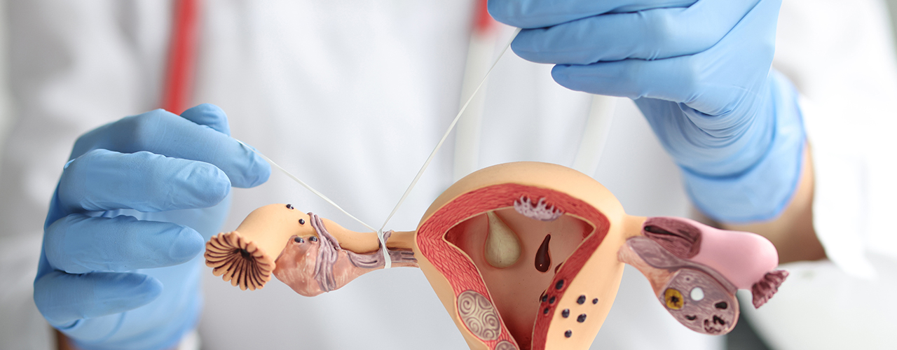
Hysterosalpingogram (HSG)

Hysterosalpingogram
For a woman to conceive it is important that all the organs of the reproductive system be healthy and functioning properly. The ovaries, uterus, fallopian tubes, and vagina need to be in a healthy state to achieve a successful pregnancy. If any of these have an abnormality, pregnancy becomes difficult. There are many diagnostic tests that are conducted on the surface of the body to identify and diagnose any issue with a particular organ, but what if the underlying cause seems to be within the organ such as a blockage in the fallopian tubes? Yes, a different type of procedure needs to be performed that allows examining within the body. Hysterosalpingogram (HSG) is one such procedure that allows the examiner to look inside the walls of the uterus and the fallopian tubes. It is a minor procedure that provides information about the contour and shape of the uterus and detects any scarring, fibroids, or polyps within the endometrial cavity. It also helps in diagnosing whether there is any blockage in the fallopian tubes.
What is HSG?
Hysterosalpingogram (HSG) is a diagnostic procedure that uses an x-ray and a special type of dye (iodine) to detect any fibroids, scar tissue, polyps, or other growths that could be causing the fallopian tubes to be blocked and preventing you from getting pregnant.
HSG step-by-step
• To prepare for the procedure you might be asked to take an over-the-counter pain medication an hour prior to performing the procedure. Depending on the condition, your doctor could also prescribe an antibiotic.
• Since it is a minor procedure, it is usually carried out in the office or at the clinic of a gynaecologist. Under a fluoroscope (an x-ray imager), a speculum is inserted into the vagina to open it and the cervix is cleaned.
• A cannula (thin tube) is then inserted into the cervix and the speculum is removed.
• The uterus is then filled with a contrast dye (iodine). The flow of the dye is then tracked by imaging with the help of a fluoroscope.
• On completion of the imaging, the procedure is completed and the cannula is removed.
The procedure does not carry any risks and is generally less painful with only mild cramping experienced during the procedure, but if there is any blockage in the fallopian tube, there is a possibility that you might experience intense pain which should subside with over-the-counter pain medications. After the completion of the procedure, the report gives a detailed picture and your doctor will be able to suggest you the appropriate treatment.